Most successful Safe & Natural Kidney Stone Treatment Without Surgery
भारत में बेस्ट किडनी स्टोन (पथरी) उपचार
Avoid Surgery! Stop suffering from pain. Get quick pain-relief & painless kidney stone removal through Dr. Bhushan’s proven homeopathic medicines. The Best Solution “Without Surgery”.
Renowned expert Dr. Kirti Bhushan offers specialized homeopathic solutions, recognized for facilitating smooth stone passage and providing immediate and complete pain relief.
Our Smiling Patients


Why Choose Us?
Quick Pain Relief !!
As fast as 15-20 minutes !!
Our Specialized Medicines Start Giving Relief from the 1st dose.
Effective Pain-Relief In A Very Short Time.
No Surgery. !! 100% Safe Cure. !!
Your Kidneys Are One of The Most important organs of your body.
There have been several instances of Kidney Damage Due to Surgical Removal of Kidney Stones That Significantly Affected Patient’s Life.
On the other hand, Our Natural Treatment is 100% Safe. Free from Painful Surgery, Cuts & Wounds and Physical & Mental Trauma.
Up to 18 mm Stones Treated Successfully !!
Check Google Reviews of Our Happy Patient’s Experience About Our Successful Treatments For Large & Small Stones
Easy & Simple Medication. !!
Our 100% Safe & Natural Treatment is based on Holistic, Simple & Easy Homeopathic Medicines.
The Scientific & Evidence Based Homeopathic System of Medicine is the Safest and Proven for Positive Treatment Response. Free from Any Side Effects.
Big Money Saving. !!
Our Pocket-friendly, Most Economical Treatment Eliminates the Need for Hospitalization and Expensive Medical Reports.
Dr. Bhushan’s Highly Specialized and Successful Kidney Stone Treatment is Available at a Fraction of Cost when Compared to the Sky-High Cost of Hospitalization, Expensive Medications and Work-Day Loss.
Based on above factors, you can very well judge which is the best option for your kidney stone removal. You can avoid surgery with Dr. Bhushan’s 100% safe & successful treatment. OR choose a more painful, high-cost surgery & risk your kidneys for entire life.
How We Treat Kidney Stones ?
Dr. Bhushan’s Kidney Stone Treatment Approach is defined in Five Elemental Steps.
1st Step
Provide Quick Pain Relief
Pain-Relief from First Dose. The powerful action of medicines helps to relieve pain normally within 15-30 minutes.
2nd Step
Pulverise & Dissolve
Forceful Medicinal Synergy Produce Lithotriptic and Anti-Lithiatic action to help dissolve Kidney Stones.
3rd Step
Clean up Kidneys & Flush-Out Small Particles
Strong diuretic action helps small stone particles to flush out with urine flow. sometimes patients don’t even feel it.
4th Step
Protect Kidneys from Damages
Nephroprotective medicines protect the kidneys from damage, improve renal function, and prevent from acute or chronic kidney disease.
5th Step
Prevents Stone Build-up Again
Preventive and therapeutic effects help to stop repeat-formation of kidney stones due to genetic & metabolic issues.
Brief Intro
Qualifications
Experience
Symptoms of Kidney Stones
Types of Kidney Stones
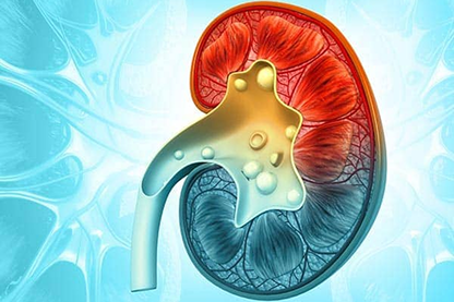
Kidney Stones
Urinary stone (Kidney stone) prevalence is estimated at 3% in all individuals, and it affects up to 12% of the population during their lifetime. Urinary stone recurrence rates approach 50% at 10 years and males have the highest incidence. Prior to the development of modern urologic techniques for treatment, mortality from untreated staghorn calculi was 27%. Currently mortality from stone disease is rare, although there is still a significant rate (28%) of renal deterioration with certain stone types.
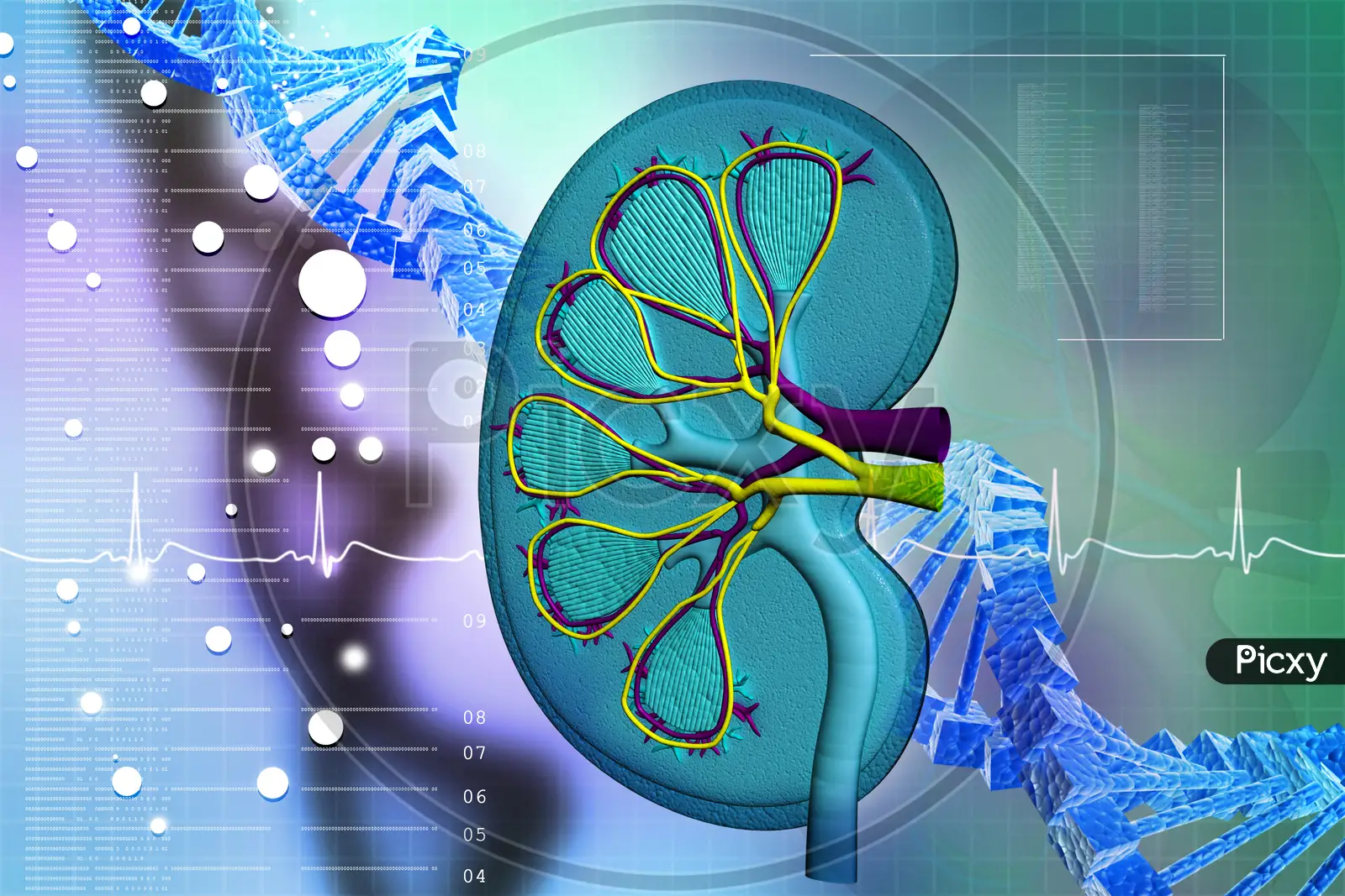
Pathophysiology
Urinary calculi may have various compositions which include, in order of decreasing frequency: calcium oxalate (monohydrate or dihydrate), uric acid, struvite (magnesium ammonium phosphate), calcium phosphate, and cystine. There are other less common stones, including xanthine and drug-related stones as well. Stones are solutes that occur in amounts too high tostay dissolved (supersaturated) in urine. As a result of supersaturation, the solutes precipitate and aggregate to form concretions or stones.
Calcium Oxalate Stones
It is thought that the majority of calcium oxalate stones form from an initial calcium phosphate concretion that originates near the renal calyx epithelium in the highly concentrated environment of the terminal collecting duct. The calcium phosphate concretion (called a Randall’s plaque) erodes through the urothelium, is exposed to urine, and forms a nidus for calcium oxalate deposition with time.
The calcium oxalate deposition grows until the stone becomes large enough to break free of its urothelial “anchor” and then may pass through the collecting system. Factors that promote calcium oxalate supersaturation (and calcium oxalate deposition) are dehydration, hypercalciuria, hyperoxaluria, hypernatrituria, and hyperuricosuria. Urinary citrate is an important inhibitor of calcium oxalate formation so hypocitraturia is a risk factor for stone formation.
Uric Acid Stones
Uric acid is a product of purine metabolism. Uric acid is 100 times more soluble at a pH > 6 compared to a pH Less than 5.5. Other than dehydration, the most common risk factor for uric acid lithiasis is persistently acidic urine including the lack of a normal postprandial alkaline tide. Likewise, patients with persistent acidosis (e.g., distal renal tubular acidosis) are also at risk for developing uric acid stones. Less commonly, gout (hyperuricemia) is associated in approximately 20% of cases with hyperuricosuria and uric acid lithiasis. Hyperuricosuria is also associated diseases such as lymphoma or leukemia that are treated with chemotherapy. With such treatment, the sudden lysis of millions of cells releases a large quantity of purines into the circulation and urine that may precipitate in the renal tubules and cause uric acid stones.
Struvite Stones
Struvite stones are caused by urinary infections with urease producing organisms, the most common being Proteus mirabilis. Less common pathogens include Klebsiella, Enterobacter, or Pseudomonas. (E. Coli is not a urease producing organism.) Urease cleaves each mole of (soluble) urea into two moles of (relatively insoluble) ammonium. As this cleavage occurs, free H+ is bound to NH3 to produce NH4, yielding OH- from water, making urine more alkaline.
Phosphate is less soluble at alkaline versus acidic pH, so phosphate precipitates onto the insoluble ammonium products, yielding magnesium ammonium phosphate. As the bacteria that produce urease remain in urine and within the stone, they continue to produce urease, and continue to cleave urea, and so large (staghorn shaped) stones may develop quite rapidly and fill the calyceal spaces of the kidney (Figure 2).
Cystine Stones
Cystine stones are produced in patients with a homozygous recessive gene defect affecting cystine transport, resulting in excessive urinary cystine. Cystine is an amino acid composed of cysteine-S-S-cysteine. The four dibasic amino acids are cystine, ornithine, lysine, and arginine, remembered by the mnemonic COLA. Normal individuals generally excrete less than 100 mg of cystine per day in the urine, whereas the majority of homozygous cystinuric patients excrete more than 200 mg per day. There are no known inhibitors of cystine.
Cystine is more soluble at a pH of 9.6 and higher compared to lower pH’s, but it is practically impossible to achieve such a high urine pH by oral alkali agents (and not without risk of calcium phosphate stone formation).
Renal Physiology With Obstruction
All stones may produce obstruction and pain. Pain is thought to occur from obstruction or renal capsular distension. With acute unilateral obstruction, in the setting of a normal contralateral kidney, the affected kidney responds in 2 phases to obstruction :
Initial 2 hours. There is increased renal pelvic pressures and renal blood flow. As renal pelvic pressure increases, glomerular filtration (GFR) decreases, as GFR represents the sum of net hydrostatic and oncotic pressures across theglomerulus.
>24 hours. Renal pelvic pressures trend down towards baseline (but remain elevated) and renal blood flow continues to diminish. If persistent, the obstruction leads to renalischemia.
Thus, obstruction from urinary stones threatens GFR, renal blood flow, and if obstruction is not relieved, renal ischemia leads to irreversible renal impairment. In general, with high-grade obstruction, renal impairment will occur within 2 weeks.
Spontaneous stone passage within the distal ureter may be facilitated with drugs that enhance expulsion.
With observation, close follow-up is needed to ensure stone passage or to follow stone growth and to watch for new infections. As stone composition is typically not known on presentation, it is important to encourage patients to collect and submit their stone for analysis, so that recurrent stone episodes may be more efficiently managed with knowledge of prior stone composition.